Research
Our vision
Rather than seeing medical imaging as just a way to generate ‘pictures’ for inspection, we use medical imaging technology as a means to observe physiological processes when and when they happen, as they happen, in the human body entirely non-invasively and in a manner that can be used to study health and disease in people and ultimately be used in clinical practice to improve patient care.
We are an interdisciplinary group united around the development and use of image analysis methods for physiological imaging. An important aspect of our work is the development of software tools that enable physiological imaging technology to be used in the fundamental understanding of human biology and physiology, and in patients as part of clinical trials and clinical practice (see our software) .
Why Physiological Imaging?
We define physiological imaging as the use of medical imaging technology to observe physiological (and biological) processes as they are happening and where they are happening. The resulting images therefore comprise spatially resolved measurements of processes happening in the body that are important to normal healthy function, and are disturbed in disease.
Physiological imaging is useful because it is
- Specific - focuses on a single physiological parameter (not a mixed contrast of a lot of things that can all vary)
- Absolute - no need for reference tissues or relative values
- Substantial - physiological effect sizes are large in pathology - this is why it is pathological because the physiology has changed
- Modifiable - physiological quantities are things we can seek to alter and are things we often want to alter with treatment
(with credit to Hanzhang Lu)
Projects
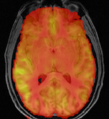
- Controlling structure induced variations in non-invasive perfusion MRI of neurodegeneration (EPSRC funded)
Perfusion imaging allows us to measure the vital role played by delivery of blood to the brain in keeping it supplied with nutrients and removal of waste. Any deviations of the blood supply from normal can be a sign of disease. In particular early and subtle changes in perfusion might mark regions of the brain which are affected by degenerative diseases such as dementia before other imaging signs become obvious. The technology exists and is increasingly widely available to image perfusion quickly and safely using Magnetic Resonance Imaging. Thus perfusion Magnetic Resonance Imaging could be a valuable tool in the understanding of dementias, as well as the diagnosis and monitoring of patients with dementia. The challenge that remains is making sufficiently specific measurements of subtle changes in blood supply that would be needed to make the technology truly useful for patients. This project addresses that problem in three ways:
- Automated removal of errors associated with imperfect measurement, for example due to motion of the patient.
- Methods to control for differences between patients due to their individual brain structure, allowing blood supply measurements to be compared between individuals or from a patient to a population of similar healthy adults. These methods remove uncertainties introduced by other differences between the brain’s of individuals that are not related to perfusion.
- Generation of personalised reference perfusion images for an individual patient against which their measured perfusion can be compared to detect changes specific to that individual.
The methods and tools that are to be generated in this project will enable perfusion Magnetic Resonance Imaging to be used more effectively in the UK-wide effort to understand dementia and in the search for new and effective treatments. Ultimately the work done in this project will enable perfusion Magnetic Resonance Imaging to become a valuable clinical tool that can be used in the diagnosis and monitoring of individual patients with dementia.
- Software
We also have a track record of releasing open-source software